News
Best Presentation Award
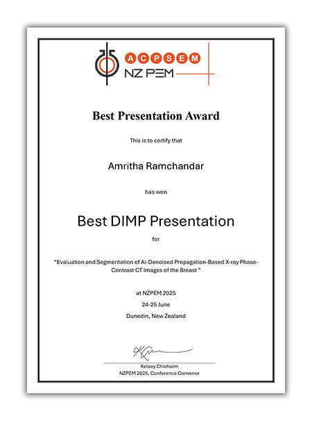
We’re proud to share a recent Best Presentation Award presented at the New Zealand Physics and Engineering in Medicine (NZPEM) Conference 2025 by Amritha Ramchandar, University of Canterbury.
This paper was co-authors by IMPACT Team Dr Ashkan Pakzad (Postdoc) and A/Prof Timur Gureyev (AI) from the University of Melbourne as part of the IMPACT NHMRC Synergy Grant.
The title of paper is “Evaluation and Segmentation of AI-Denoised Propagation-Based X-ray Phase-Contrast CT Images of the Breast”
Our Approach:
• Phase contrast imaging (PCI) to get higher SNR at lower doses
• AI to denoise low-dose phase-contrast CT images
• AI segmentation to identify adipose/glandular accurately
• Advanced imaging + AI: enhances early detection while reducing radiation dose
New Posters
🎉 We’re proud to share two posters presented at the General Breast Imaging Meeting (BIG) and the UK Imaging & Oncology Congress (UKIO) by Jenna Arbid, PhD Candidate, as part of the IMPACT NHMRC Synergy Grant.
These posters highlight our latest study on participant comfort during simulated breast propagation-based phase-contrast computed tomography (PB-CT) — a crucial step toward launching the world’s first clinical trial using PB-CT.
This cutting-edge imaging technology aims to combine high-resolution images with a more comfortable experience for patients — potentially transforming the way we approach breast cancer screening. Studies like this are helping shape protocols that don’t just meet technical standards but also prioritise the human experience.
Explore the virtual poster here
We’re delighted to announce the publication of our latest research paper in Radiography! 🎉
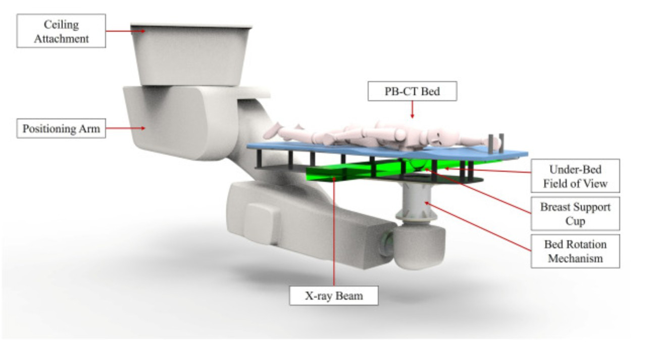
“Women’s Tolerance of Breast Propagation-based Phase-Contrast Computed Tomography (PB-CT) Positioning Procedure” 🔬
A big shoutout to our incredible IMPACT team from the University of Sydney and ANSTO:
Jenna Arbid (PhD student)
Prof Patrick Brennan, PhD Brennan (CI)
Dr Dania Abu Awwad (Lecturer)
Prof Sarah Lewis (CI)
Dr Amir Tavakoli Taba Taba (CI)
Dr Yobelli Jimenez Jimmenez (AI)
Dr Thomas Leatham (Postdoc)
Dr Daniel Hausermann (AI, ANSTO)
Dr Chris Hall (Scientist, ANSTO)
Dr Elette Engels (Researcher, ANSTO)
Led by Jenna Arbid, this study is a part of the IMPACT project funded by an NHMRC Synergy Grant.
This study supports the feasibility of PB-CT as a patient-centered imaging modality and contributes important insights as we move toward the world’s first clinical trial in breast PB-CT.
Read the full paper here
Series Presentation
Excited to share that A/Prof Kaye Morgan, CI of the IMPACT NHMRC Synergy Grant Program at Monash University, recently presented at the European Society for Molecular Imaging during their “Heart and Lung in Focus” series. Her talk, titled “Novel Approaches to X-Ray Phase and Dark-Field Imaging of the Lung”, showcased innovative techniques pushing the boundaries of lung imaging.
Watch the full presentation here
Exciting Research Breakthrough! 🚀
We’re delighted to announce the publication of our latest research paper in Scientific Reports! 🎉
“Evaluating the Feasibility of Region-of-Interest Phase-Contrast Imaging for Lung Cancer Diagnostics” 🔬
A big shoutout to our incredible IMPACT team:
From Monash University:
Lucy Costello (PhD student)
Jannis Ahlers (PhD student)
Dr Lorenzo D’Amico (Postdoc)
A/Prof Marcus Kitchen (AI)
A/Prof Kaye Morgan (CI)
Dr Daniel Hausermann (AI, ANSTO)
Dr Chris Hall (Scientist, ANSTO)
Dr Yakov Nesterets (AI, CSIRO)
Led by Lucy Costello, this study is a part of the IMPACT project funded by an NHMRC Synergy Grant and involves collaboration with top-tier institutions including Monash University, ANSTO, CSIRO and The University of Adelaide.
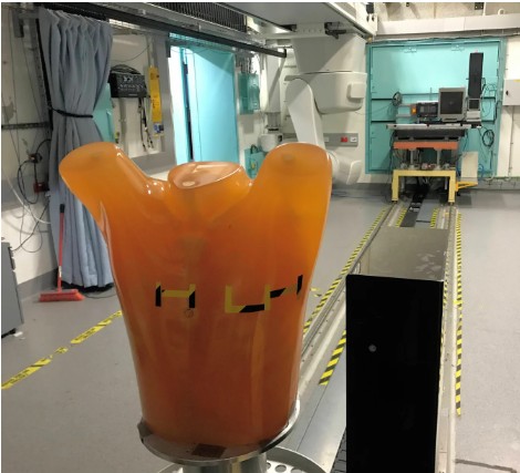
This publication to examine how phase contrast imaging can help lung and breast cancer. Using the ‘Lungman’ phantom and the Australian Synchrotron’s Imaging and Medical beamline (IMBL), pictured below, this paper tests the feasibility of ‘region-of-interest phase-contrast’ CT for lung tumour characterisation.
The researchers show that it is possible to capture a high-resolution CT of a region the size of a coin, deep within a human-sized chest, to visualise the detail of a potential lung cancer. This ‘region-of-interest’ approach is combined with propagation-based x-ray phase contrast, which provides significantly improved visualisation of the lung anatomy. Since this region-of-interest CT image is less than 2% of the area of a whole-chest CT, the x-ray beam can be significantly narrowed, which localises the associated radiation dose. We hope this novel approach can be of use in detecting and characterising lung cancer, to better inform treatment planning and improve outcomes.
Read the full paper here
🚀 IMPACT International Workshop
A Milestone in Collaboration and Innovation! 🚀

Welcome Dr Thomas Leatham
Our NHMRC IMPACT project new Postdoctoral Research Associate at the University of Sydney.
Exciting News Alert! 📣
🚀 Our latest Research Paper Published 🚀h Fellow at the University of Melbourne.
Publication of a new x-ray dark-field imaging technique
IMPACT PhD student Mr Jannis Ahlers from Monash University has published a novel dark-field imaging technique in Optica, showing that x-ray energy information can reveal where in a patient there are porous or granular structures below the resolution of the imaging system.

Welcome Dr Amir Entezam, our NHMRC IMPACT Project new Postdoctoral Research Fellow at the University of Melbourne.
Amir graduated in PhD Medical Physics (2024) from Queensland University of Technology.

Welcome, Dr Michelle K Croughan, our NHMRC IMPACT Project new Postdoctoral Research Fellow at Monash University.
Michelle completed an undergraduate degree in astrophysics in 2017, after which she transitioned to x-ray imaging research when she took on a research assistant role with Marcus Kitchen.

Welcome Dr Ashkan Pakzad, our NHMRC IMPACT Project new Postdoctoral Research Fellow at the University of Melbourne.
Ashkan graduated in MSci Medical Physics (2017) and PhD Medical Imaging (2023) from University College London.
Transforming Breast Imaging for a Brighter Future! ✨
Our Synergy Grant Research Project, funded by the National Health and Medical Research Council (NHMRC), will transform diagnosis of breast and lung cancer by establishing a path to clinical implementation of a novel low-dose, high-quality, 3D imaging technique with an emphasis on patient comfort.
Welcome, Dr Lorenzo D’Amico, Postdoctoral Research Fellow, Monash University
Dr D’Amico earned his Honours degree from the University of Trieste in 2019. Subsequently, he pursued advanced studies at the Nanotechnology PhD school of the same institution.
Our latest research paper published. 🎉Join us in celebrating this remarkable achievement! 🎉
Delighted to announce the IMPACT Research Team led by Associate Professor Timur Gureyev titled, “Young’s double-slit interference with single hard x-ray photons,” now in the Optics Express.

Jannis Ahlers at The European Synchrotron Radiation Facility
Selected as one of the 72 successful applicants from 178 applications…

Congratulations Professor Patrick Brennan!
Congratulations to Professor Patrick Brennan, Chair of Diagnostic Imaging, University of Sydney and Co-founder & CEO of DetectedX for the Award of Best University Research Commercialisation from the Australian Financial Review. Patrick is also the Chief Investigator (CIA) of the NHMRC Synergy IMPACT Program, administered by theUniversity of Sydney.

Congratulations Associate Professor Kaye Morgan!
Congratulations Dr Amir Tavakoli Taba!
Congratulations Dr Amir Tavakoli Taba (who is also the Chief Investigator of the NHMRC Synergy IMPACT Program) on initiating such a worthwhile STEM program collaborated between USYD, University of Southern Queensland and Catholic Education Diocese of Bathurst, and it now being recognised by media, including the 7 News. Dr Taba stated that “aligned with the USYD’s “Sydney in 2023” strategy, we’re creating opportunities and bridging educational disparities, making a significant impact in the space of STEM education.”
Congratulations to the research team members who successfully applied for academic promotion in 2023.
Level D – Associate Professor
Dr Kaye Morgan, School of Physics and Astronomy, Monash University
Level C – Senior Lecturer
Dr Amir Tavakoli Taba, School of Health Sciences, University of Sydney
Dr Nicola Giannotti, School of Health Sciences, University of Sydney
Dr Yobelli Jimenez, School of Health Sciences, University of Sydney
The sneaky breast cancer I almost missed — and how other women can look out for it – ABC News
Medical experts, including Professor Mary Rickard, a member of our IMPACT Research Program Clinical Implementation Expert Committee (CIEC), provided insights on women dealing with lobular breast cancer and offered guidance on detection, aligning with the objectives of our IMPACT Research Program. She recommends additional imaging techniques, such as contrast mammograms and MRIs.
